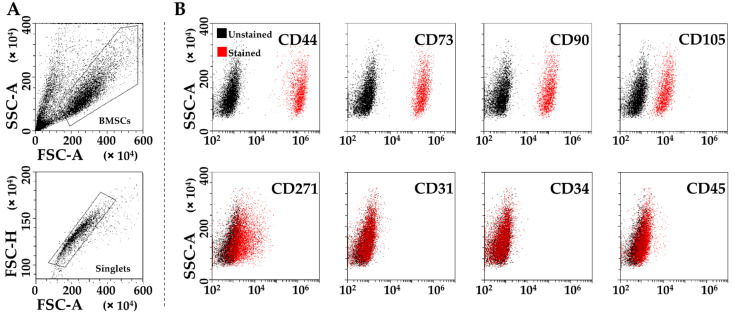
Figure 1.
Flow cytometry analysis of SF-treated BMSCs. (A), after exclusion of debris (upper panel), single cells were identified (lower panel). (B), staining of single cells for general mesenchymal (CD44, CD73, CD90 and CD105, positive), BMSC-specific (CD271, positive) and hemato-endothelial markers (CD31, CD34, and CD45, negative). Representative plots are shown.

