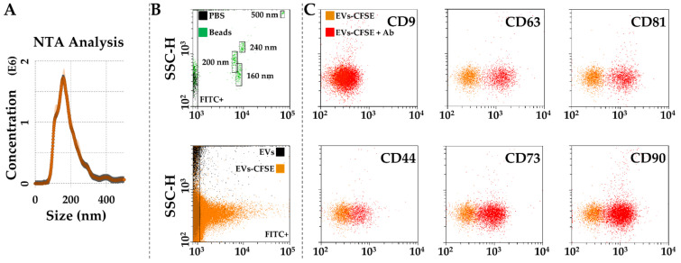
Figure 5.
Characterization of SF-treated BMSC-EVs. (A), EVs size analysis from NTA data. (B), flow cytometer was first calibrated to score FITC-fluorescent particles of nanometer scale (upper panel, starting from 160 nm). EVs were CFSE stained to allow their identification and gating in the FITC channel (lower panel). (C), after gating, CFSE+ EVs showed positive staining for extracellular vesicle defining molecules CD63 and CD81, and MSC markers CD44, CD73 and CD90. CD9, another EV postulated marker, was barely detectable. Representative cytograms are presented.

