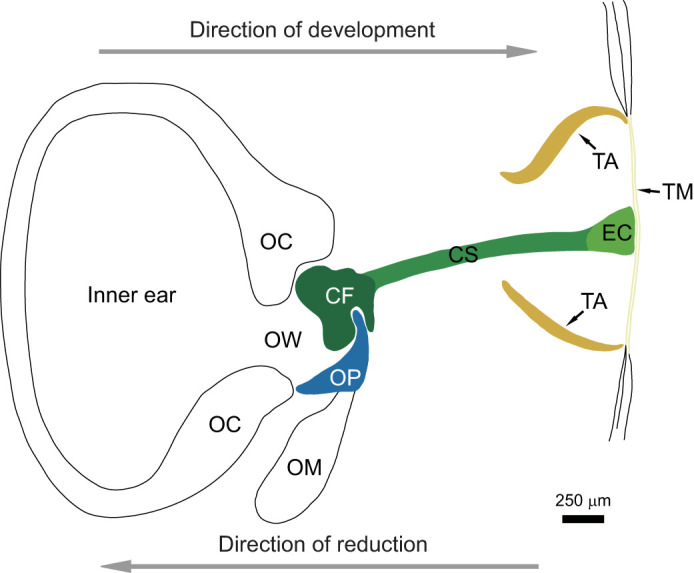
Fig. 1.

Diagrammatic sketch of a fully functional tympanic middle ear at the final stage of metamorphosis in Rana pipiens. During secondary reduction of middle ear structures, the most peripheral structures are lost before the more medial structures (arrow showing direction of reduction). CF, columellar footplate; CS, columellar shaft; EC, extra-columella; FO, fenestra ovalis, oval window; OC, otic capsule; OM, opercularis muscle; OP, operculum; OW, oval window; TA, tympanic annulus; TM, tympanic membrane. The operculum is formed first (blue); the columella is then formed in three parts: columellar footplate (first), then columellar shaft and extra-columella (last) (dark green to light green). The tympanic annulus (TA) is formed before the tympanic membrane (TM). Modified from Hetherington (1987).
