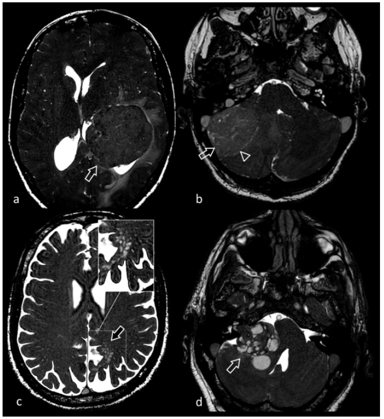
Figure 10.
(a) Axial 3D CISS image shows a large mass (arrow) in the left lateral ventricle. 3D CISS accurately depicts the endoventricular location of the mass and helps characterize its relationship with the surrounding brain tissue. Histological findings were in keeping with intraventricular fibrous meningioma. (b) 32-year-old female patient with a large lesion (arrow) in the posterior fossa, initially suspected for a cerebellar neoplasm. 3D CISS optimally demonstrates a subtle hyperintense rim between the mass and the cerebellar hemisphere (CSF cleft sign, arrowhead), suggestive for the extra-axial location of the tumor. After brain surgery, final diagnosis was extra-axial desmoplastic medulloblastoma of the right cerebellopontine angle. (c) Incidental subcortical and cortical lesion (arrow) in the left parietal lobe, hyperintense in T2-TSE and FLAIR sequences, devoid of contrast enhancement or diffusion restriction, and without signs of mass effect. 3D CISS shows with high definition that the lesion is mainly made of a cluster of multiple well-defined «bubbles». Findings were consistent with a multinodular and vacuolating neuronal tumor. (d) 62-year-old woman. Axial 3D CISS image shows a well-defined lesion, with solid and cystic components, in the right cerebellopontine angle that partially extends into the internal acoustic canal and causes compression of the antero-lateral portion of the pons, which was later diagnosed as vestibular schwannoma histopathologically.

