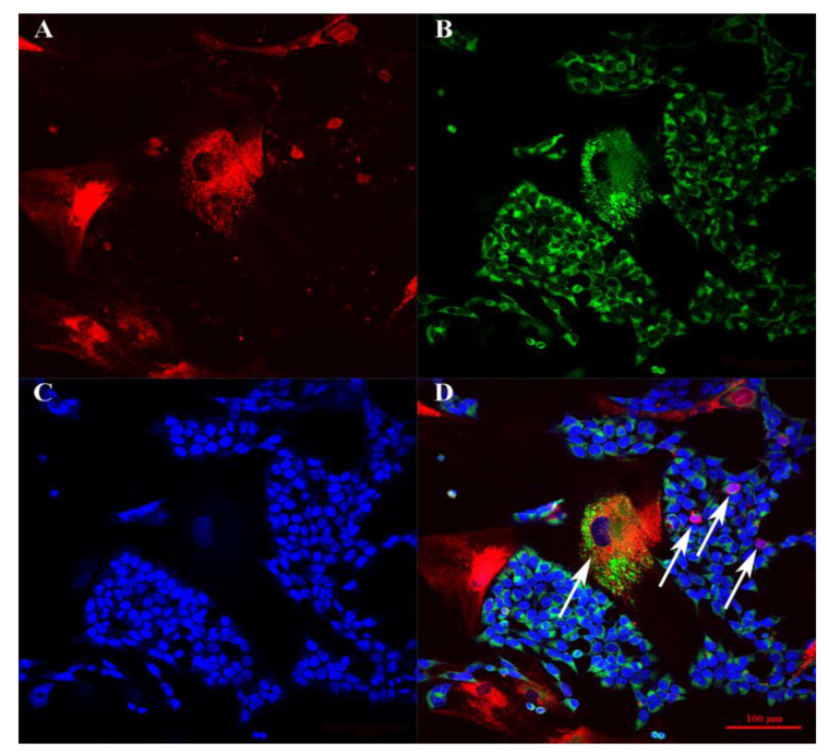
Figure 4.
The confocal microscopy BM-MSCs and SH-SY5Y cells on glasses. (A) Channel filtered for the emission spectrum of Alexa Fluor 647. (B) Channel filtered to gather fluorescence emission from Alexa Fluor 488. (C) Channel filtered for the emission spectrum of Alexa Fluor 460. (D) Merge. Arrows indicate the cells that have mixed colors as a result of membrane and cytoplasmic exchange between cells.

