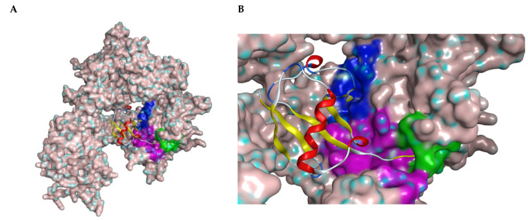
Figure 1.
(A) Structure of USP7 catalytic domain in complex with ubiquitin (PDB ID: 1NBF). The molecular surface was added. The ribbon model represents ubiquitin molecule. (B) Closer look of the binding region of USP7 inhibitors. The green surface depicts the catalytic center; the purple surface depicts the cleft between the Thumb and Palm domains; the blue surface depicts the allosteric binding site.

