Figure 7.
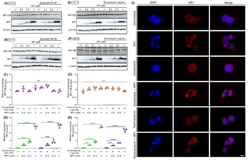
Expression of Neuropeptide Y and NPY-Y2R receptor in SH-SY5Y cells. (A,B) Western immunoblot analysis illustrates the presence of NPY-Y2R in all indicated groups (Control, Control + NPY, glutamate, and glutamate + NPY) and dose-dependent increase in the NPY expression with NPY treatment at 12 and 24 h, respectively. (C,D) Densitometric quantification of NPY-Y2R blot intensities represent that its expression was not affected by either the insults (glutamate or tunicamycin) or the NPY treatment at 12 h (F(3,16)= 5.461 in Figure (C) and F(3,16) = 1.265 in Figure (D), ns represents non-significance, n = 3). (E,F) Western immunoblot analysis illustrates the presence of NPY-Y2R in all indicated groups (Control, Control + NPY, tunicamycin, and tunicamycin + NPY) and dose-dependent increase in the NPY expression with NPY treatment at 12 and 24 h, respectively. (G,H) Densitometric quantification of NPY blot intensities showed a progressive dose-dependent significant increase in its expression with NPY treatment (F(3,16)= 264.8 in Figure (G,F) (3,16) = 697.9 in Figure (H), ** p < 0.01, *** p < 0.001, **** p < 0.0001 represents significance between control and NPY group (green), ** p < 0.01, *** p < 0.001, **** p < 0.0001 represents significance between insults (glutamate or tunicamycin) and insults + NPY (purple), n = 3) and no expression in untreated cells or in cells exposed to only insults (glutamate or tunicamycin). Each band intensity was normalized to the respective band intensity of β-actin. (I) Immunofluorescence images (representative) of treated SH-SY5Y cells showing the expression of NPY. After 12 h of treatment, cells were fixed and immunostained with an anti-NPY antibody (red). Nuclei were stained with DAPI (blue). Scale bar = 10 μm.

