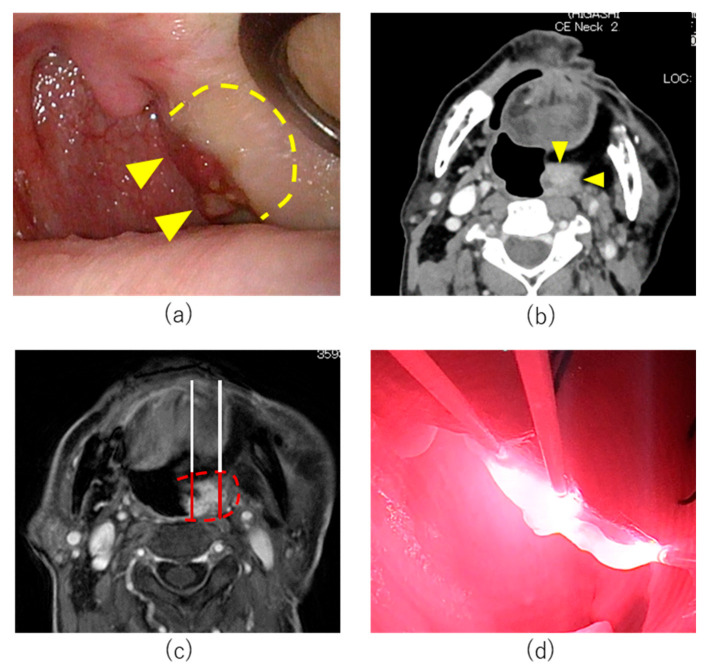
Figure 2.
Treatment course of Case 1. (a) Pretreatment endoscopic findings: Left oropharyngeal tumor (arrowheads) and tumor induration (dotted line). (b) Axial section of Gd-enhanced MRI T1 weighted image. (c) Treatment plan of cycle 1 HN-PIT treatment. Planned illumination area (dotted line). (d) Intraoperative image of HN-PIT in Cycle 1. Near-infrared laser was illuminated using 20 mm cylindrical diffusers.

