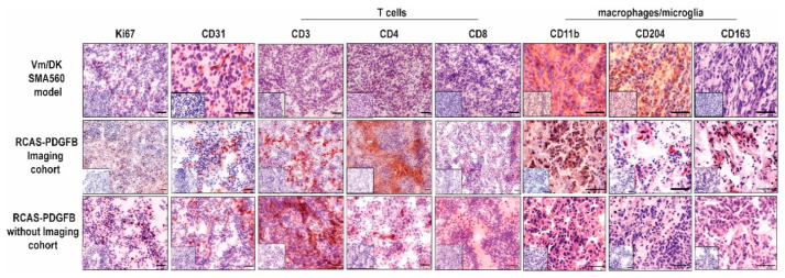
Figure 4.
Immunohistochemical comparison of established T cell, microglia/macrophages, and vascularisation markers as indicated above. Representative IHC staining patterns of tumor tissues of either VM/Dk mice treated with an isotype control antibody (MOPC-21), as described in Przystal et al., or transgenic mice after the implantation of RCAS-PDGFB transfected DF1 cells. Small inserts show staining control without the application of primary antibody. Scale bars are 100 µm.

