Figure 1.
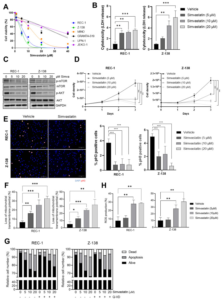
MCL cytotoxicity, proliferation impairment and cell death induced by simvastatin. (A) Cell viability of MCL cell lines in the presence or absence of different doses of simvastatin; (B) cytotoxicity rates assessed by LDH release of MCL cell lines in presence or absence of 5 μM, 10 μM and 20 μM simvastatin; (C) Western blot evaluation of phospho-AKT and phospho-mTOR levels after the treatment with 5 μM, 10 μM and 20 μM simvastatin, or vehicle. Full western blot images can be seen in Figure S4. (D) Proliferation rate assessed by MCL cell counting after the treatment with increasing doses of simvastatin; (E) proliferation rate assessed by phospho-Histone 3 (red) immunofluorescence of MCL cell lines in the presence or absence of 5 μM, 10 μM or 20 μM simvastatin. Nuclei were counterstained with DAPI (blue); (F) mitochondrial transmembrane potential (ΔΨm) after cell treatment with 5 μM, 10 μM or 20 μM simvastatin or vehicle; (G) apoptosis rate measured by AnnexinV-FITC/PI staining after cell treatment with 5 μM, 10 μM or 20 μM simvastatin or vehicle, previously treated or not with 10 μM of the pan-caspase inhibitor, Q-VD-OPh hydrate; (H) ROS generation measured by DHE (dihydroethidium) staining in MCL cells lines treated with 5 μM, 10 μM or 20 μM simvastatin, or vehicle. Values are expressed as mean ± SEM. * p < 0.05, ** p < 0.01, *** p < 0.001, when compared to control group.

