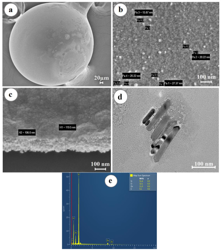
Figure 3.
Morphological analysis of the surface of the ball resonator optical fiber coated with a ZnO layer: (a) SEM image of the optical fiber sensor showing its spherical shape and surface coating; (b) SEM image demonstrating the grain structure of the ZnO layer; (c) ZnO layer thickness on the surface of the optical fiber biosensor; (d) TEM image of ZnO; (e) EDS—surface analysis of the ball resonator optical fibers demonstrating the presence of Zn, O, C, and Si elements.

