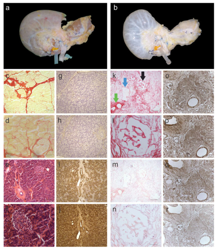
Figure 1.
Characterization of decellularized human pancreas tissue. (a) before and (b) after decellularization. Yellow/pink tissue is a representative of cellular material, which is the colour of fresh pancreata, whereas the translucent-white color is a representative of decellularized tissue. Histological images of native pancreas stained with Picro-Sirius Red (SR) and imaged at (c) 10× and (d) 40× magnification. SR stains cellular material yellow and collagen fibres red. Histological images of native pancreas stained with H&E and imaged at (e) 10× and (f) 40× magnification. H&E stains nuclear material blue and the ECM red. Immunohistology images of native pancreas stained for (g) collagen I, (h) collagen III, (i) fibronectin and (j) laminin. Histological images of decellularized pancreas stained with SR and imaged at (k) 10× and (l) 40× magnification. SR staining showed no evidence of cellular material. Histological images of decellularized pancreas stained with H&E and imaged at (m) 10× and (n) 40× magnification. H&E staining showed no evidence of nuclear material. Immunohistology images of decellularized pancreas stained for (o) collagen I, (p) collagen III, (q) fibronectin and (r) laminin. The black arrow is pointing at an islet of Langerhans, the green arrow is pointing at an exocrine ECM duct and the blue arrow is pointing at a blood vessel, which were well maintained in decellularized pancreata. Scale bars for (c,k,e,m) are 200 µm, for (d,f,l,n) are 50 µm and for (g–j,o–r) are 100 µm. (n = 6 pancreata).

