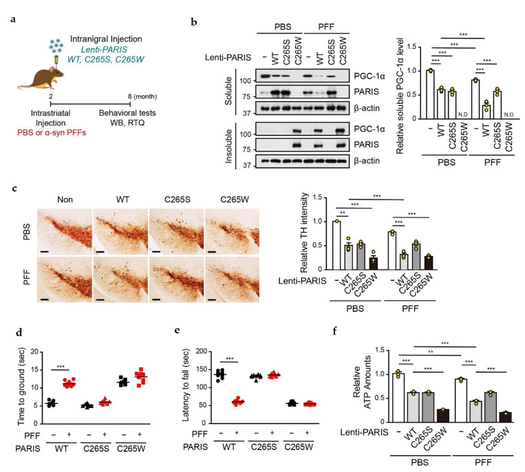
Figure 6.
Loss of functional PGC-1α due to α-syn PFFs is SNO-PARIS-dependent. (a) Study timeline for intrastriatal α-syn PFFs injection and intranigral injection of lentiviruses (pLenti-GFP, pLenti-PARIS WT, pLenti-PARIS C265S, and pLenti-PARIS C265W). Biochemical and behavioral testing was performed as indicated in the timeline, and all mice were sacrificed after behavioral experiments. (b) PARIS and PGC-1α levels in RIPA-soluble and RIPA-insoluble fractions of the SN region of α-syn PFFs/lentiviruses injected mice. Right panel—relative PGC-1α level in the soluble fraction normalized to β-actin level. n = 3. Two-way ANOVA followed by Bonferroni’s post-test, *** p < 0.001. N.D, not detected. (c) Representative TH staining of the midbrain sections from the SN region of α-syn PFFs/lentiviruses-injected mice. Scale bar = 200 μm. Right panel—relative TH intensity was measured by using the ImageJ software. n = 4. Two-way ANOVA, followed by Bonferroni’s post-test, ** p < 0.01, *** p < 0.001. Pole test (d) and rotarod test (e) of α-syn PFFs/lentiviruses-injected mice at the age of 8 months. n = 8–9 per group. Two-way ANOVA followed by Bonferroni’s post-test, *** p < 0.001. (f) Relative ATP levels in the SN region of α-syn PFFs/lentiviruses-injected mice using the ENLITEN luciferase assay. n = 3. Two-way ANOVA followed by Bonferroni’s post-test, ** p < 0.01, *** p < 0.001.

