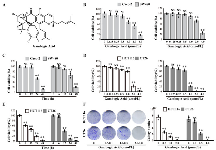
Figure 1.
GA decreases Caco-2, SW480, HCT116, and CT26 cell viability in dose- and time-dependent manners. (A) Chemical structure of GA. (B) Caco-2, SW480, (D) HCT116, and CT26 cells were treated with GA (0–4 μmol/L) for 24 h, and cell viability was analysed by the CCK8 assay. (C) Caco-2 and SW480 cells were treated with GA (2 μmol/L) for different times, and cell viability was analysed by the CCK8 assay. (E) HCT116 and CT26 cells were treated with GA (2 μmol/L and 1 μmol/L, respectively) for different times, and cell viability was analysed by the CCK8 assay. (F) HCT116 and CT26 cells were treated with the indicated concentrations of GA and stained with crystal violet. The cells were exposed to 0.1% DMSO as control. All experiments were repeated three times, and results were expressed as mean ± SD. NS, not significant, ** p < 0.01 vs. control.

