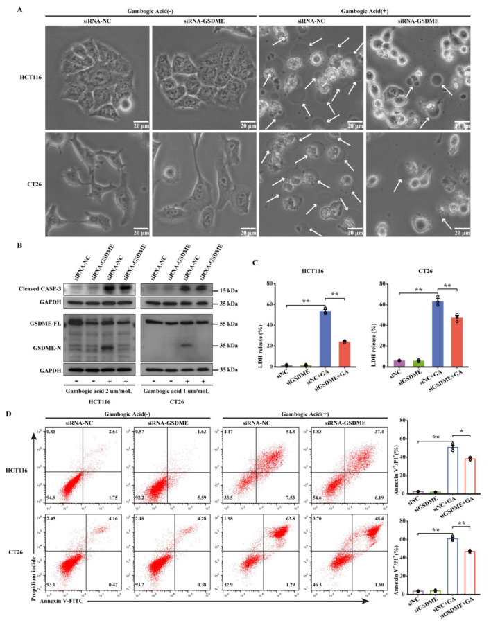
Figure 5.
GSDME mediated pyroptosis of CRC cells in response to GA treatment. HCT116 and CT26 cells were transfected with siGSDME or siNC and then treated with GA (2 μmol/L or 1 μmol/L) for 12 h. (A) SiNC- or siGSDME-infected HCT116 and CT26 cells were treated with GA, and representative bright-field images were obtained. The white arrows point to pyroptotic cells. (B) Expression of GSDME and cleaved caspase-3 in siNC- or siGSDME-transfected HCT116 and CT26 cells treated with GA analysed by western blotting. The whole western blots were shown in Figure S4. (C) The release of LDH and (D) flow cytometric analysis were performed after treatment with GA. The cells were exposed to 0.1% DMSO as control. All experiments were repeated three times and results were expressed as mean ± SD. NC, negative control. * p < 0.05 vs. control, ** p < 0.01 vs. control.

