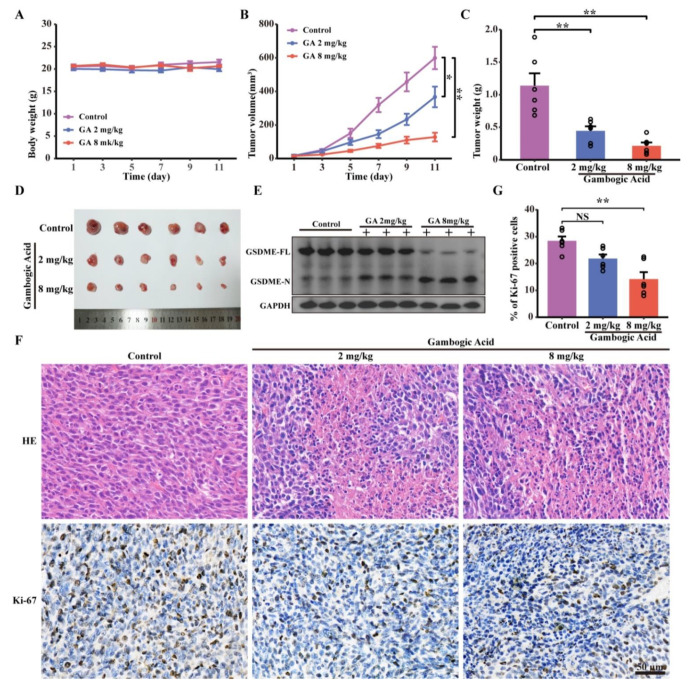
Figure 7.
GA inhibited the tumor growth and triggered pyroptosis of CRC cells in vivo. CT26 cells were inoculated into BALB/c mice to establish the murine xenograft model. (A) Change curves of mice weight after GA treatments (n = 6). (B) Tumor volume, (C) tumor weight, (D) photographs of isolated tumor after GA treatment. (E) Western blotting analyses of GSDME-FL and GSDME-N expression in treated tumor tissues. The whole western blots were shown in Figure S6. (F) H&E staining and Ki-67 immunohistochemistry analysis of tumor tissues after GA treatments. (G) Quantitation of Ki-67 staining from immunohistochemical analysis. NS, not significant, * p < 0.05 vs. control, ** p < 0.01 vs. control.

