Figure 1.
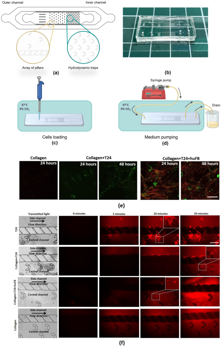
Modeling of DOX delivery to cancer cells in the presence of collagen using MFCs. (a) Geometry of the MFC. (b) Resulting MFC. (c) Scheme of the experiment with microfluidic chips. Cells loading into the MFC channel and further storage under CO2 and 37 °C for cell culture adhesion. (d) Medium pumping by a syringe pump connected to the MFC with a tubing system that leads the flow to the drain. (e) Representative SHG images of collagen seeded with T24 cancer cells or co-culture of T24 and human fibroblasts HuFb or without any cells. Red—SHG signal from collagen, green—autofluorescence of cells. Scale bar: 50 µm, applicable to all images. (f) Images of MFCs in transmitted light and representative time-lapse images of DOX fluorescence in different models. DOX was added in the concentration of 50 µg/mL (IC50). The areas of DOX uptake into T24 cells are shown in the dashed squares and enlarged in the right upper corner of the images. Red—fluorescence of DOX. Scale bar: 400 µm, applicable to all images.

