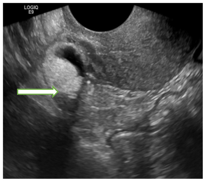
Figure 2.
Transvaginal ultrasound image of the pelvis in a 55 years old lady shows a right adnexal 1.8 × 1.8 × 1.9 cm mass with mixed echogenicities (fat, calcification, and soft tissue) consistent with a dermoid cyst. The lesion remained stable in subsequent follow up studies confirming its benign nature.

