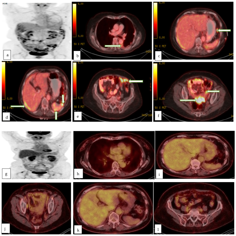
Figure 10.
Maximum intensity projection (a), and multiple axial color fused images (b–f) of PET CT in a 78 year old lady with recently diagnosed left ovarian serous adenocarcinoma for staging, show left pleural effusion with hypermetabolic nodule/implant (proven to be malignant effusion) with hypermetabolic implants in the subdiaphragmatic (c), perihepatic and perisplenic regions (d), infracolic omentum (e) and recto-vaginal pouch (f) in keeping with metastasis. Follow up maximum intensity projection (g), and multiple axial color fused images (h–l) of PET CT in the same patient after 3 months of chemotherapy, shows excellent post-treatment response with resolution of the peritoneal metastases as well as the prior left pleural effusion.

