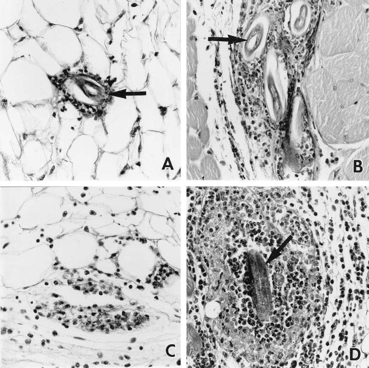
FIG. 4.
Representative photomicrographs of leukocyte infiltrates and larvae at the site of inoculation in nontransgenic (A and C) and Tg5C2 IL-5 transgenic (B and D) mice. Tissues were recovered 2 (A and B) and 12 (C and D) h p.i. At 2 h p.i., inflammatory infiltrates had formed around larvae (arrows) in both nontransgenic and transgenic mice, with both larvae and leukocytes more numerous in the latter. At 12 h p.i., leukocyte infiltrates had often largely resolved in nontransgenic mice and larvae were absent (C), whereas eosinophil-rich infiltrates had developed further around larvae still retained at the site of inoculation in transgenic mice (D). H&E-stained 5-μm sections; ×320 magnification.

