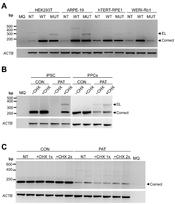
Figure 2.
Analysis of RPE65 gene expression by RT-PCR in different cellular models. (A) Midigene splicing assays in ARPE-19, WERI-Rb1, hTERT-RPE1 and HEK293T. Representative electrophoresis gel (n = 2) of the RT-PCR product after amplifying exon 1 to 3 of RPE65 comparing wild-type (WT) and mutant (MUT) expression vectors. NT: non-transfected. (B) Representative gel of the RT-PCR of control (CON) and patient-derived (PAT) iPSC and photoreceptor precursor cells (PPCs). Cells were grown in absence (−CHX) or presence of cycloheximide (+CHX). (C) Representative gel of the RT-PCR of control (CON) and patient-derived (PAT) RPE cells. The cells are untreated (NT), treated with the normal amount of cycloheximide (+CHX 1×) or the double amount of cycloheximide (+CHX 2×). ACTB amplification was used as loading control. MQ: milliQ water; EL: exon-elongation; correct: wild-type transcript; bp: base-pairs.

