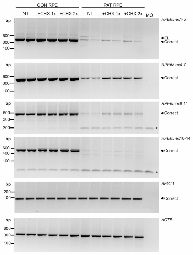
Figure 3.
RT-PCR analysis in iPSC-derived RPE cells. Different regions of RPE65 were amplified in non-treated RPE cells (NT) and RPE cells upon treating the cells with the normal dose (+CHX 1×) or double dose (+CHX 2×) of cycloheximide for both the control (CON) and patient (PAT RPE cells. All amplicons showed RPE65 reduction in PAT RPE. * indicates primer dimers. BEST1 was used as RPE marker detection and ACTB was used as a loading control. MQ: milliQ water; EL: exon-elongation; correct: wild-type transcript; bp: base-pairs.

