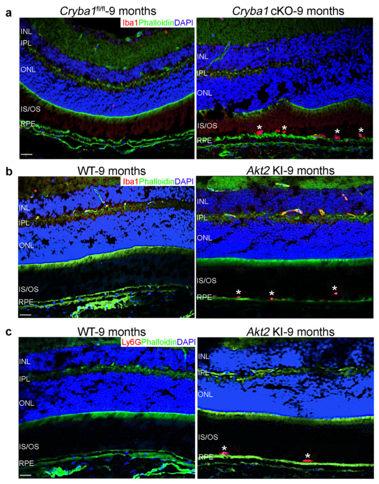
Figure 1.
Infiltration of microglia and neutrophils into the SRS of aged Cryba1 cKO and Akt2 KI mice. (a) Immunofluorescence studies showing accumulation of Iba1-positive microglial cells (Iba1: Red, asterisks) in the SRS of retina sections from 9 month old Cryba1 cKO mice, but not in age-matched Cryba1fl/fl retina sections. Scale bar = 100 µm. (b,c) Retina sections from 9 month old Akt2 KI mice showing subretinal infiltration of microglia (Iba1: Red, asterisks in (b), and neutrophils (Ly6G: Red, asterisks in (c). Green: Phalloidin, Blue: DAPI; nucleus. Scale bar = 100 µm. n = 3.

