Figure 4.
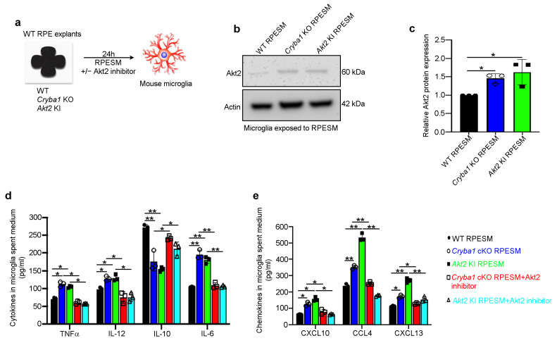
RPE-mediated microglial activation is regulated by Akt2. (a) Schematic showing experimental design; RPE spent medium (RPESM) harvested from 24 h RPE explant cultures from aged WT, Cryba1 KO and Akt2 KI mice was used to culture mouse microglia for 24 h with or without 5 nM Akt2 inhibitor. (b) Western blot and (c) densitometry showing that Akt2 is upregulated in microglial cells following exposure to RPESM from Cryba1 KO and Akt2 KI RPE explant cultures, relative to WT RPESM-exposed microglia. (d) Cytometry bead array analysis revealed significant upregulation of M1 mediators TNFα, IL-12, and IL-6 and downregulation of the M2 mediator IL-10 in MSM from Cryba1 KO and Akt2 KI RPESM-exposed microglia relative to WT RPESM-exposed cells. This trend indicates a transition to the pro-inflammatory M1 state in these microglia. (e) Chemokines such as CXCL10, CCL4, and CXCL13 were also increased in the spent medium from Cryba1 KO and Akt2 KI RPESM-exposed microglia, compared to controls. Surprisingly, adding Akt2 inhibitor to the microglia culture medium rescued the levels of these pro-inflammatory mediators to near control values (d,e). n = 3. * p < 0.05, ** p < 0.01.

