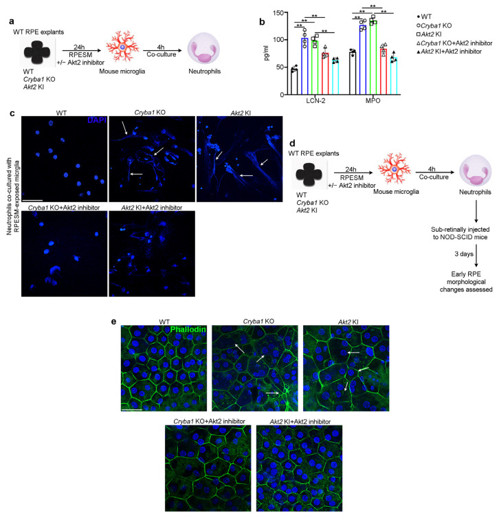
Figure 6.
Pro-inflammatory microglial cells trigger neutrophil activation and thereby induce RPE changes. (a) Schematic showing experimental design; RPE spent medium (RPESM) harvested from RPE explant cultures (24 h culture) from aged WT, Cryba1 KO and Akt2 KI mice were used to culture mouse microglia for 24 h. Neutrophils were then co-cultured with the RPESM-exposed microglia with or without 5 nM Akt2 inhibitor. (b) ELISA showing significant increase in neutrophil activation markers LCN-2 and MPO in neutrophils co-cultured with microglia cells which were exposed to RPESM from Cryba1 KO or Akt2 KI RPE explant cultures, relative to neutrophils co-cultured with WT RPESM-exposed microglia. Akt2 inhibitor treatment to Cryba1 KO and Akt2 KI RPESM treated microglia before neutrophil co-culture, reduced the levels of LCN-2 and MPO in these cells. n = 4. (c) DAPI (blue) staining showing extended nuclear projections (NET formation) in neutrophils co-cultured with Cryba1 KO or Akt2 KI RPESM (arrows); Akt2 inhibitor treatment to the microglia prior to co-culturing with neutrophils markedly reduced this effect. n = 3. Scale bar = 50 µm. (d) Schematic showing study design for subretinal injection of neutrophils into NOD-SCID mice (as described in (a)). After 3 days the extent of early RPE changes was assessed. (e) Immunofluorescence studies on RPE flatmounts from NOD- SCID mice showing significant alterations of normal honeycomb such as morphology and enlarged cells (arrows in (e)) in the central region of the flatmounts from eyes injected with activated neutrophils (co-cultured with microglia which were treated with Akt2 KI or Cryba1 KO RPESM), compared to control. Akt2 inhibition caused marked reduction in the morphological alterations in the RPE (e). n = 5. Scale bar = 50 µm. ** p < 0.01.

