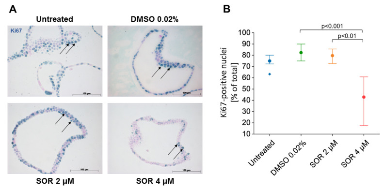
Figure 3.
Sorafenib inhibits cell proliferation in iCCAOs in a concentration-dependent manner. (A) iCCAOs were treated for 48 h as indicated with the vehicle control (DMSO 0.02%), or with 2 and 4 µM sorafenib. Proliferating cells were identified by immunohistochemical detection of the nuclear antigen Ki67 (arrows). (B) For quantitative evaluation, positive cells in each 10 visual fields were detected using ImageJ and expressed as percentage number of total cells. Data were not normally distributed as verified by the Shapiro–Wilk test. The Kruskal–Wallis test and post-hoc Bonferroni analysis of significance were performed. Statistical analysis was accomplished using SPSS. Horizontal lines indicate the P-level of significance.

