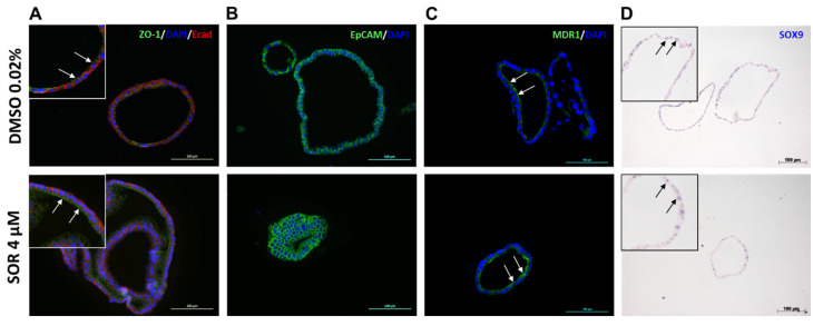
Figure 4.
Sorafenib treatment does seemingly not affect protein expression of epithelial markers and proliferation marker SOX9 in iCCAOs. (A) iCCAOs were treated for 48 h as indicated either with the vehicle control (DMSO 0.02%), or with 4 µM sorafenib, resp. Fluorescent immunohistochemistry revealed apical and basolateral expression of ZO-1 (green) and E-cadherin (red), resp. Nuclei are labeled with DAPI (blue). Insets show digital magnifications (orig. magnification: 20×) of selected areas of the pictures behind. Arrows indicate luminal expression of ZO-1. (B) iCCAOs were treated for 48 h as indicated. Fluorescent immunohistochemistry showed membranous expression of EpCAM (green). Nuclei are labeled with DAPI (blue). (C) iCCAOs were treated for 48 h as indicated. Fluorescent immunohistochemistry revealed apical expression of MDR1 (green, white arrows). (D) iCCAOs were treated for 96 h as indicated. SOX9 expression was localized in the nuclei (insets, black arrows in digital magnifications (orig. magnification: 10×) of selected areas of the pictures behind) as shown by non-fluorescent immunohistochemistry.

