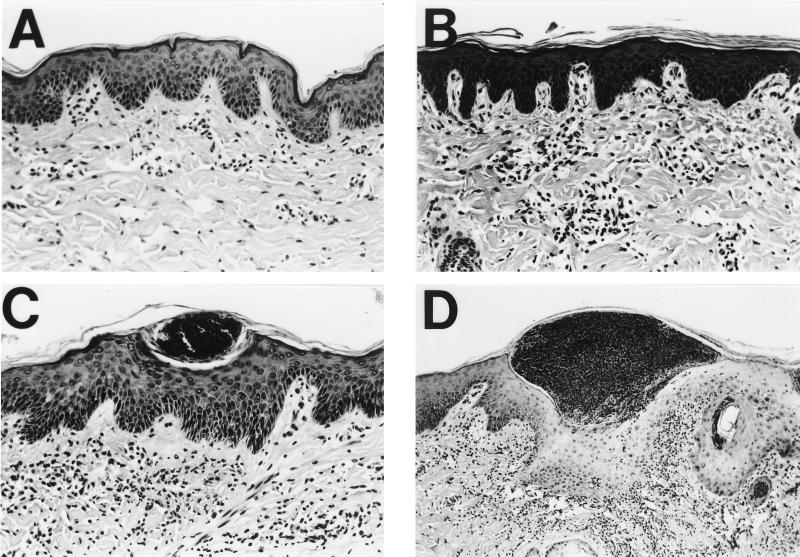
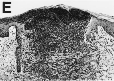
FIG. 2.
Hematoxylin- and eosin-stained cross sections of pig skin inoculated with H. ducreyi, demonstrating method of scoring lesion severity. (A) Skin of normal appearance, score = 1; (B) Presence of dermal perivascular and interstitial mononuclear cell infiltrate, score = 2; (C) Presence of intraepidermal pustule consisting of neutrophils, fibrin, and necrotic debris, score = 3; (D) Epidermal pustule accompanied by keratinocyte cytopathology and diffuse mononuclear and polymorphonuclear dermal infiltrate, score = 4; (E) Ulceration or epidermal necrosis and dermal erosion accompanied by confluence of immune cells, score = 5. Magnifications, ×103 for panels A to C and ×52 for panels D and E.


