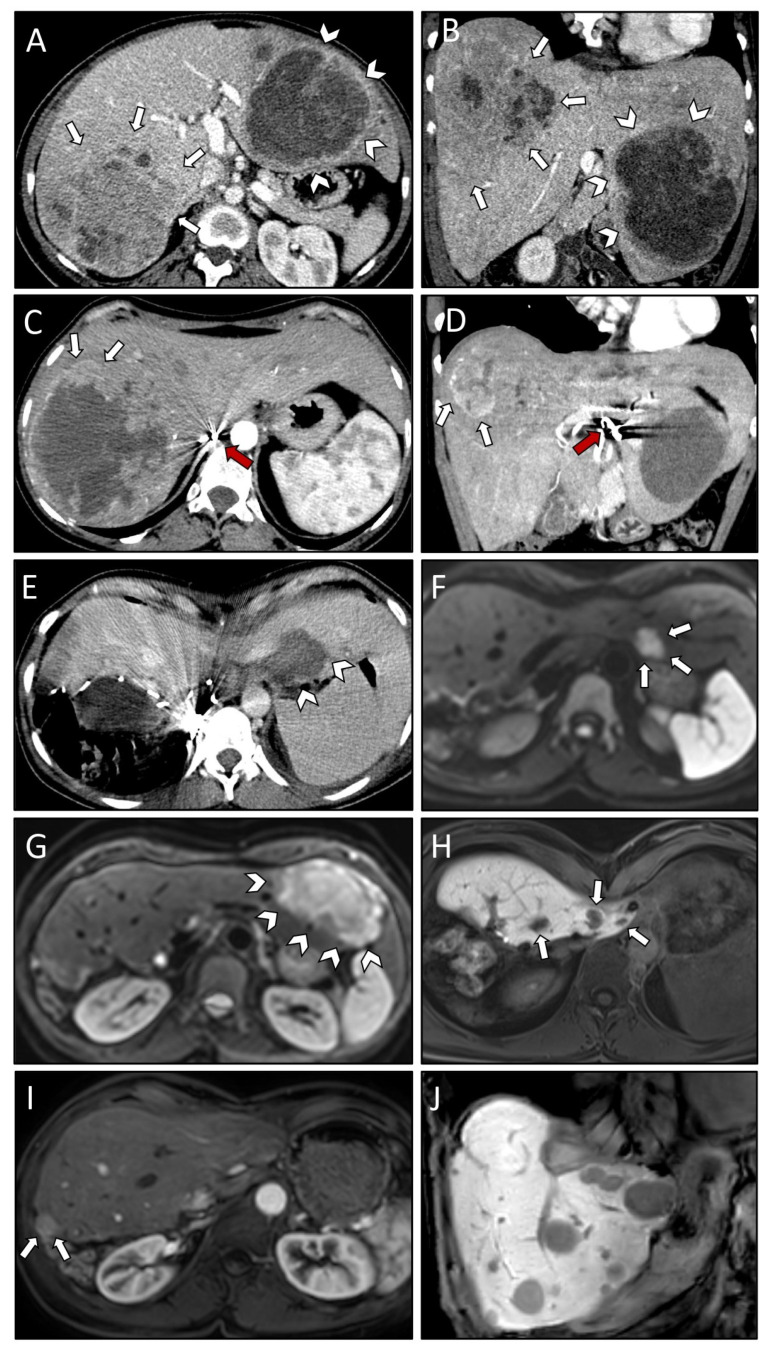
Figure 2.
Axial (A) and coronal plane (B) of initial CT scans demonstrating very large SFT/HPC liver metastases in both right (white arrows) and left (white arrowheads) liver lobe. CT scan in axial (C) and coronal plane (D) after three TAE sessions revealing insufficient devascularization of the right liver lobe lesion anteriorly (white arrows). Note the beam hardening artifacts (red arrows) in the hepatic artery. Post-interventional CT scan (E) after first SRFA with laparoscopic liver packing (protection of cardio-esophageal region) showing coagulation zone (white arrowheads) with no residual vital tumor tissue visible. Contrast-enhanced MRI scan four months after first SRFA revealing a new metastasis in liver segment II (white arrows; (F)) and local recurrence in segment III following TAE (white arrowheads; (G)). MRI scan depicting three of six new liver lesions in segment IVa (white arrows; (H)) after resection of left lateral segments, all of them successfully treated with SRFA. MRI scans 15 months after the last intervention showing local recurrence at the resection margin (white arrows; (I)) and multiple new liver metastases (J).

