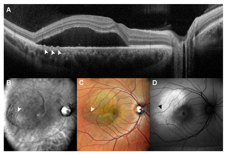
Figure 1.
Patient with central serous chorioretinopathy assesed by multimodal imaging (I). A patient with central serous chorioretinopathy is assessed by spectral domain optical coherence tomography (SD-OCT) (A), scanning laser ophthalmoscopy (SLO) retromode imaging (B), SLO color fundus imaging (C) and fundus autofluorescence (FAF) (D). In this case, we have an extended amout of SRF, which creates hyper-autofluorescence signalling in FAF, covering a mild RPE dystrophy temporally to fovea (arrows, highlighting RPE dystrophy sseen at SD-OCT). SD-OCT was able to identify RPE elevations, while SLO retromode was able to identify RPE dystrophy despite the presence of fluid thanks to retroillumination.

