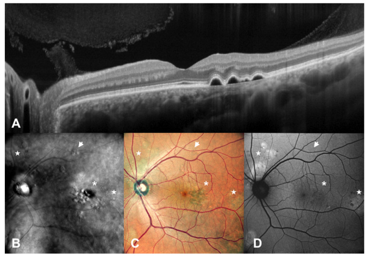
Figure 3.
Patient with central serous chorioretinopathy assesed by multimodal imaging (III). A patient with central serous chorioretinopathy is assessed by spectral domain optical coherence tomography (SD-OCT) (A), scanning laser ophthalmoscopy (SLO) retromode imaging (B), SLO color fundus imaging (C) and fundus autofluorescence (FAF) (D). In this case, we have the presence of RPE mottling (arrows) and pigmented epithelium detachments (asterisks) which are detectable with SLO Retromode imaging, while they are not seen with FAF. Flat areas of RPE dystrophy and areas of ellipsoid zone damage, exposing RPE fluorophores, can be clearly identified with FAF, while they are difficult to spot with SLO retromode imaging. (stars).

