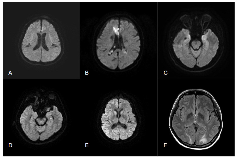
Figure 3.
(A) Status epilepticus presents a left frontal cortical hyperintensity in DWI; (B) acute ischemic infarction in right ACA territory with DWI hyperintensity; (C) autoimmune encephalitis presents with bilateral hyperintensity in DWI and T2 FLAIR; (D) dot-like hyperintensity of DWI in TGA; (E) diffuse gyriform hyperintensity of DWI in cerebral cortex in sCJD; (F) bilateral hyperintensity of T2 FLAIR in white matter regions without abnormalities in DWI in PRES.

