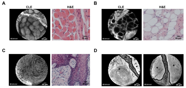
Figure 3.
Comparison between CLE and H&E morphology of tissue in the head and neck area. Confocal laser endomicroscopy (CLE) images (left picture, fresh frozen samples) and H&E-stained slides from the respective FFPE tissue are shown for skeletal muscle of the soft palate (A), adipose tissue of the cheek (B), a nonkeratinizing squamous cell carcinoma of the tonsil (C) and a keratinizing squamous cell carcinoma of the tongue (D). As shown in (D), tumor borders (black line), as well as tumor localization (black star), can clearly be identified using CLE technology. CLE and H&E images are shown in 40× magnification.

