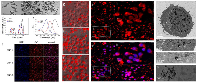
Figure 1.
The transmission electron microscope imaging of the three kinds of GNRs: (a) GNR-L, (b) GNR-S, (c) GNR-C. (d) Particle size analysis of the three kinds of GNRs. (e) UV analysis of the three kinds of GNRs. Representative images from the HCS after GNR (GNR-L, GNR-S, GNR-C) exposure to Hep G2 cells for 12 h. (f) Cell nucleus (blue) and GNRs (red). Images were acquired with an High Content screening (HCS). HCS images of Hep G2 cells treated with GNR-L for multiple time points, (g) 0 h, (h) 12 h, (i) 24 h. Confocal images of Hep G2 cells treated with GNR-L for 12 h, (j) Cy5-labeled GNR-L, (k) Cy5-labeled GNR-L and DAPI labeled Hep G2 cells. Part II in the figure is the enlarged result of part I in the figure. Biological transmission electron microscope imaging of Hep G2 cells treated with GNRs, (l) 2 μm, (m) 1 μm, (n) 0.5 μm, (o) 500 nm.

