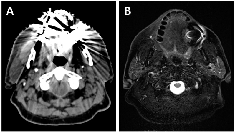
Figure 2.
A 70-year-old patient with dental amalgam planned for concurrent chemoradiotherapy for a cT3N2b squamous cell carcinoma of the right base of tongue. The planning CT (A) shows poor image quality with important artifacts which could easily obscure oral or oropharyngeal pathology. The planning T2-weighted MRI (B) also shows artifacts due to dental amalgam, but the image quality remains sufficient for adequate radiological evaluation of most of the oral cavity and oropharynx.

