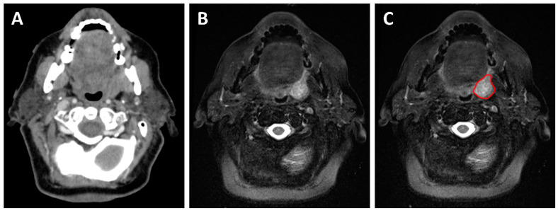
Figure 4.
A 73-year-old patient with a cT2N1 p16-positive squamous cell carcinoma of the left tonsil planned for concurrent chemoradiotherapy. The planning contrast-enhanced CT (A) shows an asymmetrical left tonsil with poorly defined margins. The planning T2-weighted MRI (B,C) allows better visualization of the tumor margin and aids in the delineation of the gross tumor volume during radiation treatment planning (red contour).

