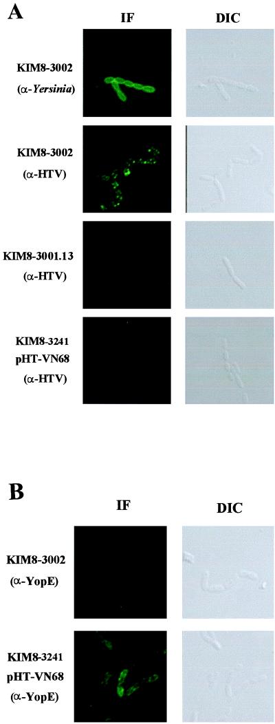
FIG. 7.
LcrV can localize to the surface of Y. pestis. Surface deposition of LcrV and YopE was tested in Y. pestis strains lacking pPCP1. Parent Y. pestis (KIM8-3002), secretion-negative Y. pestis (yscC KIM8-3001.12), and the lcrV-null strain (KIM8-3241) complemented with pHT-VN68 were fixed on coverslips after incubation for 2 h at 37°C in RPMI. Bacteria were stained with α-Yersinia or α-HTV (A) or α-YopE (B). The respective proteins were then visualized by treatment with Oregon green-conjugated secondary antibody followed by confocal laser-scanning microscopy. Immunofluorescence (IF) confocal and differential interference contrast (DIC) images are shown.

