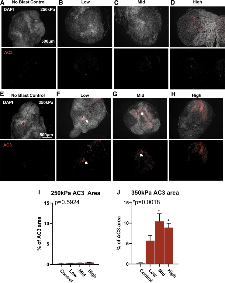
FIG. 6.
Higher amplitude blast increases cellular damage. (A) Representative image of no blast control organoid. Top, individual cell nuclei stained with 4′,6-diamindino-2-phenylindole. Bottom, immunohistochemistry for activated caspase 3 (AC3) to indicate cellular damage and programmed cell death. (B–D) Representative histology of an organoid exposed to Low, Mid, and High at 250 kPa amplitude pressure, respectively. (E–H) Representative histology of organoids exposed to Low, Mid, and High, at 350k Pa. (I) Quantification of AC3 area from organoids exposed to all frequencies at 250 kPa. (J) Quantification of AC3 area from organoids exposed to all frequencies at 350 kPa. All data presented as percent (%) area of AC3 to organoid area. Error bars presented as standard error of the mean. *p < 0.05, n = 6-12 organoids per group. All statistics calculated using one-way analysis of variance. Color image is available online.

