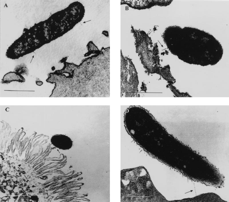
FIG. 6.
Transmission electron micrographs (thin sections, ruthenium red stain) of A. veronii biovar sobria adhering to HEp-2 cells; (A and B) and freshly isolated human enterocytes (C and D). Note the gap between the organism and the cell membrane as well as pili (arrows) extending into space or interacting with the apical brush border. Bars, 0.5 μm (A), 0.5 μm (B), 1 μm (C), and 0.2 μm (D).

