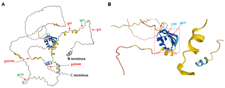
Figure 2.
Predicted three-dimensional (3D) structure of human POLDIP3 using the AlphaFold Protein Structure Database (https://alphafold.ebi.ac.uk/, accessed on 19 October 2022). (A) The 3D structure of the full-length protein of human POLDIP3. (B) The 3D structure of the RRM domain of human POLDIP3 is highlighted with a dashed red square. The two residues flanking the five APIM motifs, arginine-53 and lysine-124, are shown in GREEN colored fonts. The two residues flanking the RRM domain, threonine-280 and asparagine-351 are shown in BLUE colored fonts. The serine residues (serine-42, -63, -383/385, and -405/406) that can be phosphorylated are shown in RED colored fonts.

