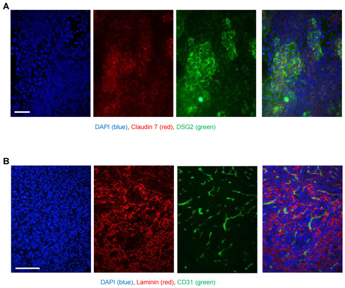
Figure 4.
Immunofluorescence analysis of TC1-DSG2 tumor sections. Tumors were harvested when they reached a volume of 500–600 mm3, embedded in OCT, and sectioned. (A) Sections were stained for (human) DSG2 (green) and the epithelial marker claudin 7 (red). Visible are nests of human DSG2-postitive tumor cells embedded in tumor stroma cells (mouse-derived). (B) Sections were stained for the extracellular matrix/stroma protein (red) and the endothelial/blood vessel cell marker CD31 (green). The images show a vascularized tumor with typical stroma components.

