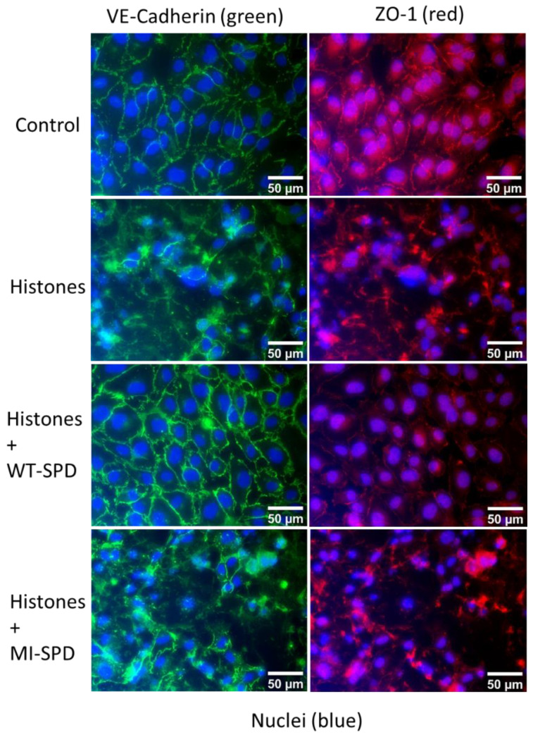
Figure 3.
Effect of SPD–FSAP on the pattern of junctional proteins. HUVEC were treated with 50 μg/mL histones and/or 10 μg/mL WT–SPD–FSAP for 24 h before fixation. MI–SPD–FSAP was used as a negative control. Immunofluorescent staining of the junction protein VE-cadherin (green), ZO-1 (red), and nuclei (blue). Relative IgG was used as control. Representative images from 3 independent experiments.

