
Figure. Tmem100, a lung-specific endothelium gene.
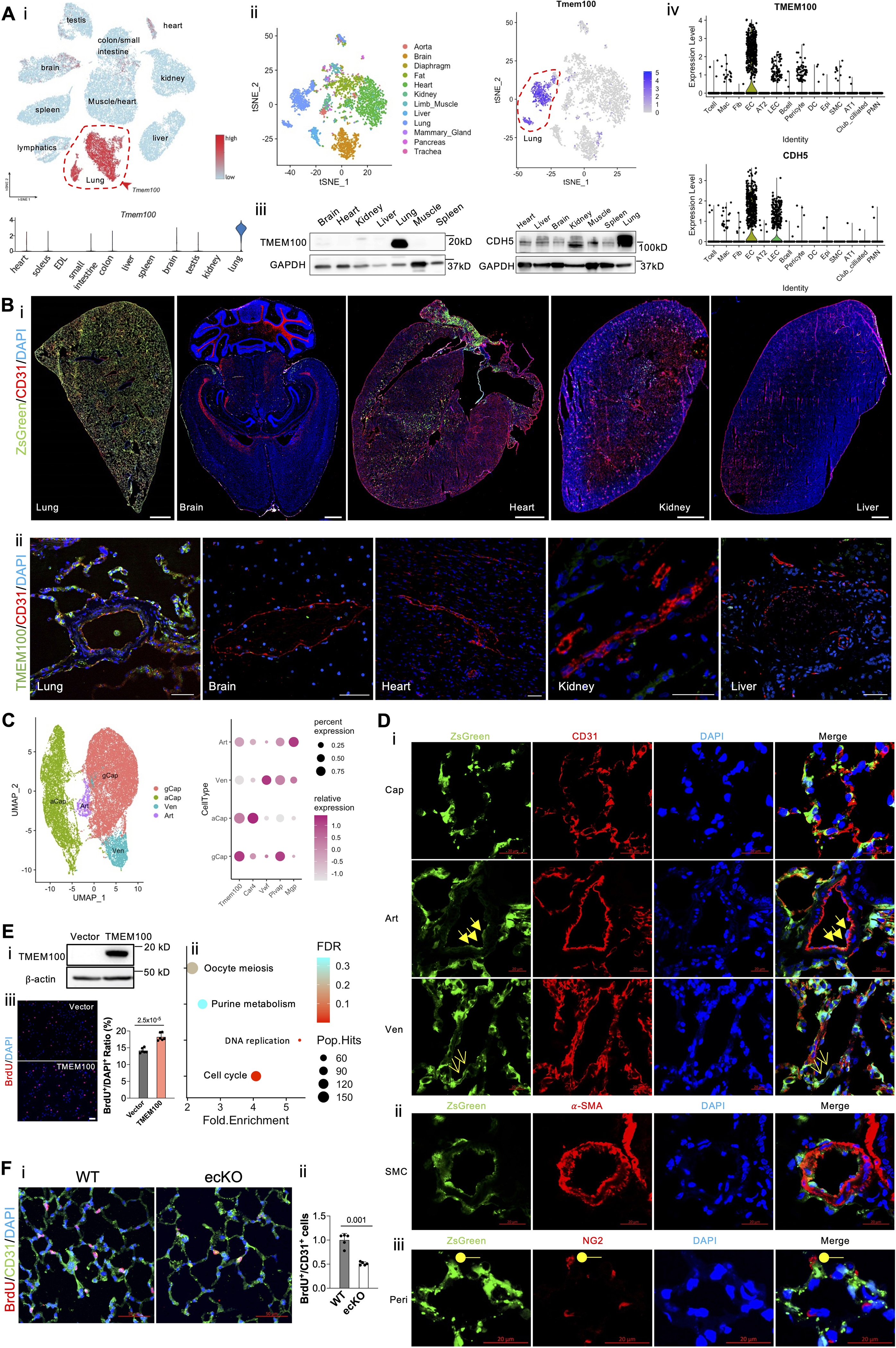
(A, i). A representative tSNE plot and a violin plot of murine ECs scRNA-seq data showing the lung EC-specific expression of Tmem100 in comparison to other organs’ ECs. (A, ii). A representative tSNE plot of extracted EC transcriptomic analysis of Tabula Muris scRNA-seq dataset demonstrating unique Tmem100 expression pattern in lung ECs. (A, iii). Western Blotting demonstrated Tmem100 protein selectively expressed in the mice lung. GADPH was used as a loading control. (A, iv). A representative Violin Plot showing that TMEM100 was selectively expressed in the lung ECs similar to classical EC marker CDH5 in human. (B, i). A lineage tracing strategy using Tmem100ZsG mice demonstrated that Tmem100 (ZsGreen+) was enriched in the lung ECs (CD31+) but not the ECs in other organs such as brain, heart, kidney, and liver. Both male and female Tmem100ZsG mice at the age of 7 weeks were injected intraperitoneally with tamoxifen daily for 5 days (20mg/kg, daily) and rested for ~10 days. Scale bar: 1 mm. (B, ii). Immunostaining demonstrated TMEM100 is selectively expressed in human lung ECs. Scale bar: 50 μm (C). A UMAP plot showing the different subtypes of lung ECs based on the murine EC dataset, and Tmem100 highly expressed in general capillary EC (gCap), aerocytes (aCap), and lowly expresses in arterial (Art) and venous (Ven) ECs. (D, i) Immunostaining demonstrated that ZsGreen+ (Tmem100+) was strongly expressed in the capillary ECs and weakly expressed in the arterial and venous ECs. Cap, capillary EC; Art, arterial EC (triangle arrows); Ven, venous EC (arrows). (D, ii) ZsGreen+ (Tmem100+) was not expressed in SMCs (α-SMA+). (D, iii) A few NG2+ cells (pericytes, Peri, round shape) expressed ZsGreen (Tmem100). Scale bar: 20 μm. (E). TMEM100 overexpression (E, i) in HPMVECs induced enrichment of pathways related to Cell Cycle, DNA Replication (E, ii) based on the upregulated genes induced by TMEM100 overexpression. Three replicates were pooled in the equal amount for RNA-seq analysis. (E, iii). TMEM100 overexpression promoted EC proliferation assessed by BrdU assay. At 48 hours post-lentiviruses infection, HPMVECs were starved in serum/growth factors free medium for 12 hours. BrdU (10μM) was added in the medium at 4 hours prior to cells harvest. BrdU was stained with anti-BrdU antibodies. Scale bar: 100 μm. N=6 (biological replicates). Student t test. (F, i, ii). Genetical deletion of Tmem100 in mice impaired EC proliferation in vivo during inflammatory lung injury. Tmem100f/f (WT) and Tmem100EndoSCL-CreERT2 (ecKO) mice were treated with LPS (5mg/kg) intraperitoneally. Mice were injected BrdU (25 mg/kg) intraperitoneally daily for two days. Lung tissue were collected at 72 hours post-LPS treatment. BrdU was stained with anti-BrdU antibodies. N=5 (biological replicates). Welch’s t-test. Statistics. (GraphPad Prism 9.0): based on additional literature support from similar studies, our samples fit normal distribution. Parametric test: parametric test with the unpaired 2-tailed Student t test for equal variance and Welch’s t-test for unequal variance (2 groups). Mean±SD.