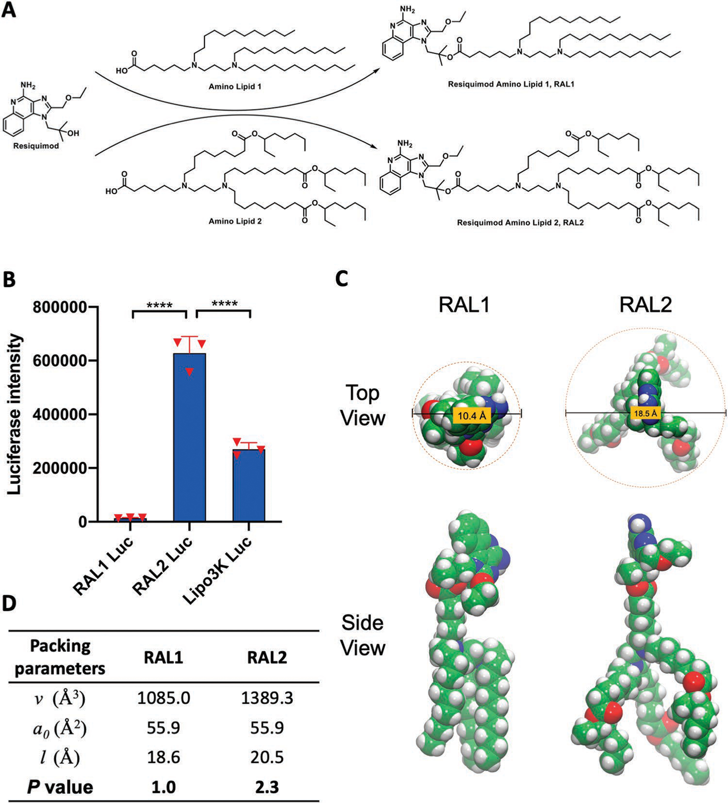
Figure 2.

Structure and characterization of RAL-LNPs. A) The synthetic routes to RAL1 and RAL2. B) Delivery of Luc mRNA in JAWS II cells. Data in D are presented as the mean ± standard deviation (S.D.) (n = 3). C) Structural illustration of RAL1 and RAL2. D) Critical packing parameters calculated for RAL1 and RAL2 structures sampled from molecular dynamics simulation. Statistical significance in (B) was analyzed by one-way ANOVA with Dunnett’s multiple comparison test. ****p < 0.0001.
