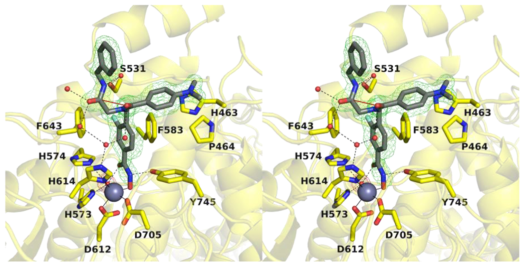
Figure 3.

Stereoview of a Polder omit map (contoured at 4.0σ) depicting the monodentate binding of 10h (dark gray) in the active site of HDAC6 chain D (yellow) (PDB 7U8Z). The catalytic Zn2+ ion is shown as a grey sphere, and water molecules are shown as small red spheres. Metal coordination, hydrogen bond, and n→π* interactions are shown as solid, dashed black, and solid red lines, respectively.
