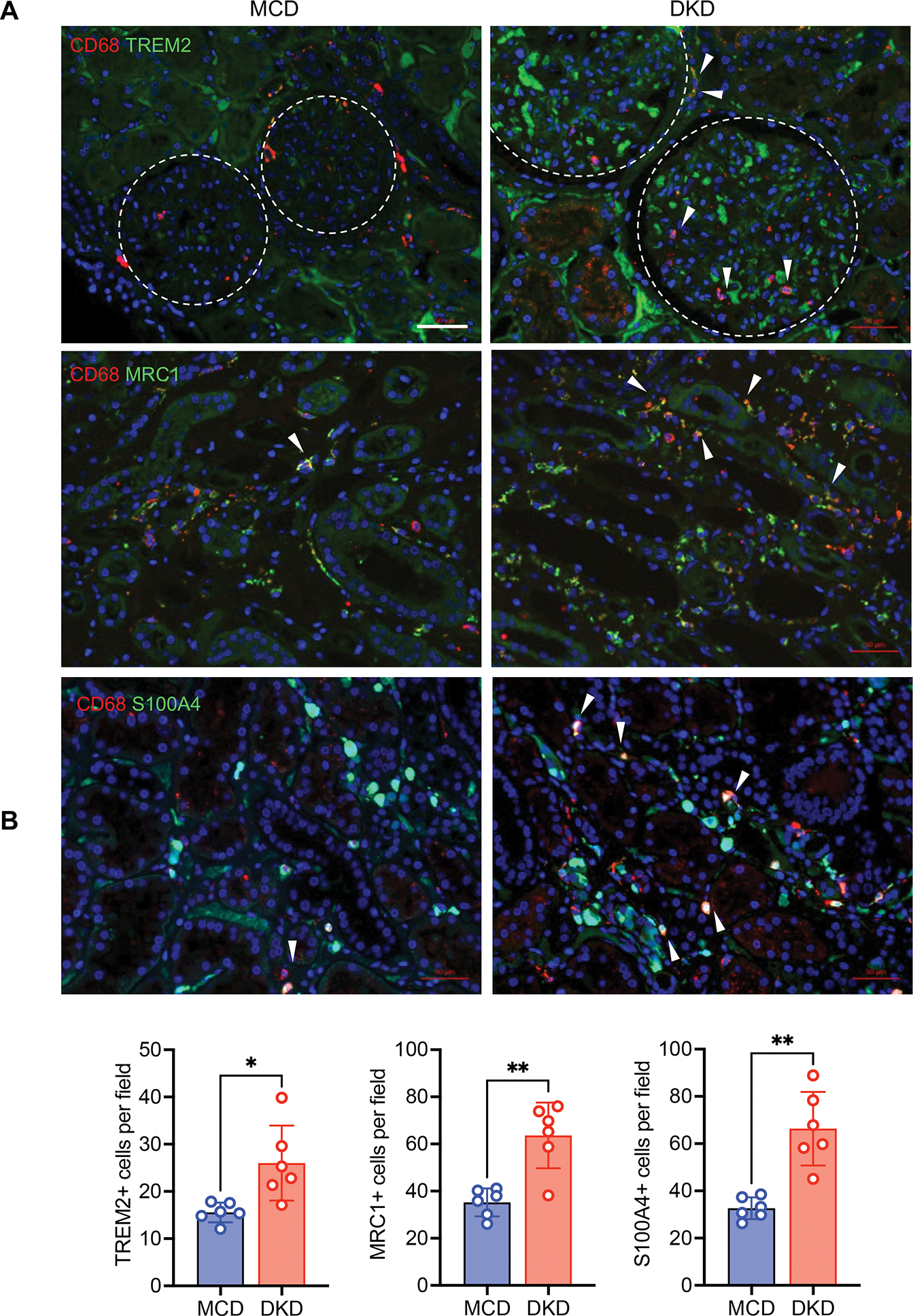
Figure 7. Increased select macrophage subsets in human DKD.

(A) Representative immunofluorescence images of TREM2, MRC1, and S100A4 in biopsy samples of DKD and minimal change disease (MCD) (n=6 DKD and 6 MCD samples). Macrophages are co-immunostained with CD68 and DNA is counterstained with DAPI. Arrows indicate double positive staining macrophages. Arrows show examples of double-positive stained macrophages. Dotted line circle glomerulus boundaries. Scale bars, 50μM. (B) Quantification of immunostaining for MRC1, TREM2, or S100A4 per field (n=6 samples per group, 5–8 fields scored per sample). ***P <0.001 between groups by Welch’s t-test.
