Figure 12.

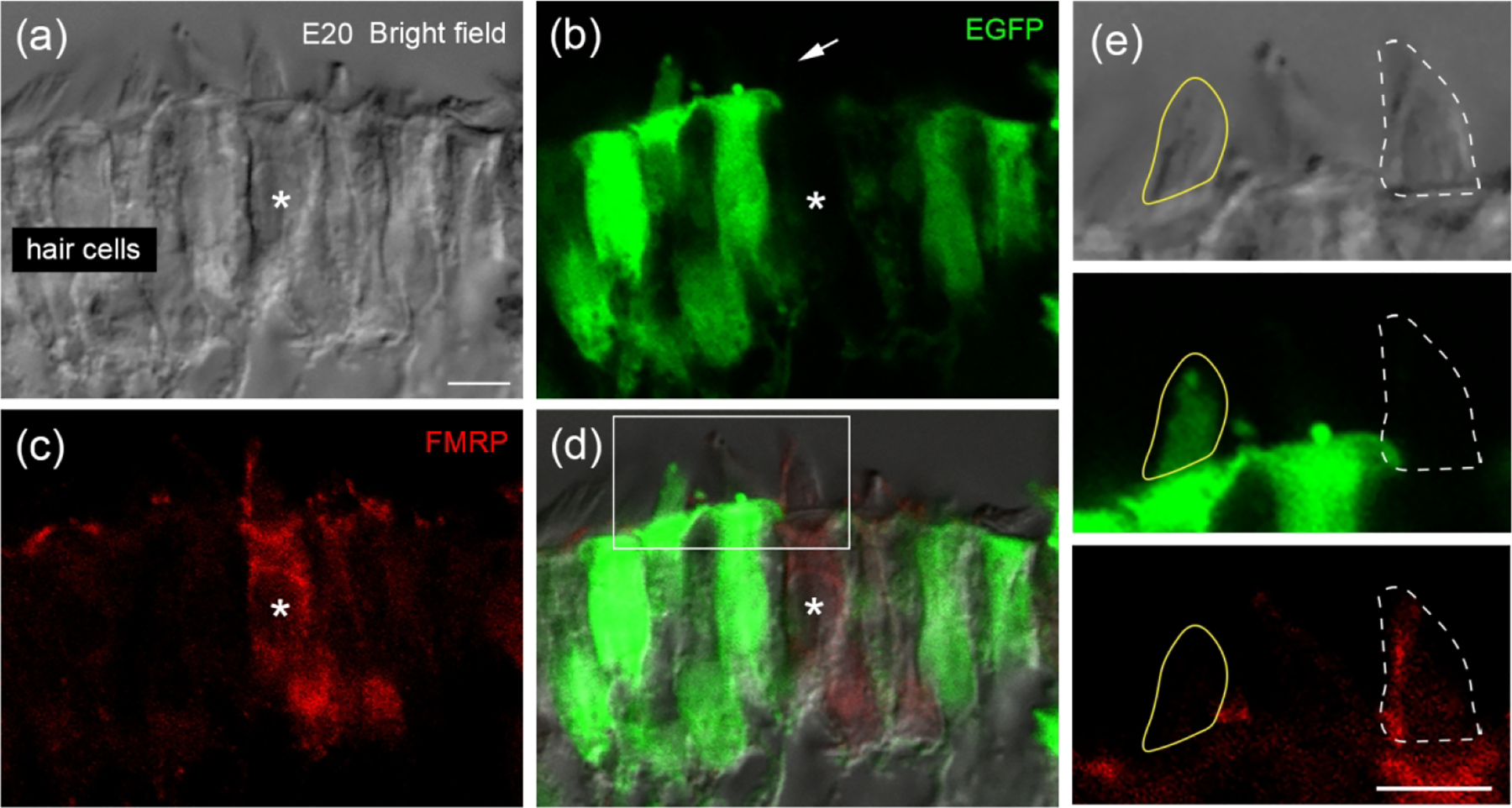
FMRP localization in hair bundles of chicken hair cells. Chicken otocysts were transfected with Fmr1 shRNA-EGFP at E2 via in ovo electroporation. FMRP immunostaining was performed at E20 on cochlea cross sections. (a) Hair cells under bright field with phase contrast. (b) Fmr1 shRNA-EGFP (green). (c) FMRP_PA8263 immunoreactivity (red). (d) Merged image. Asterisks indicate a non-transfected hair cell showing FMRP-immunoreactive hair bundle (arrow). (e) Enlarged images of the box in d. Yellow circle outlines the hair bundle of a transfected hair cell (EGFP-positive) without detectable FMRP immunoreactivity. White dashed circle outlines the hair bundle of a non-transfected hair cell (EGFP-negative) showing distinct FMRP immunoreactivity. Scale bars = 5 μm in a, applies to a–d; 5 μm in e.
