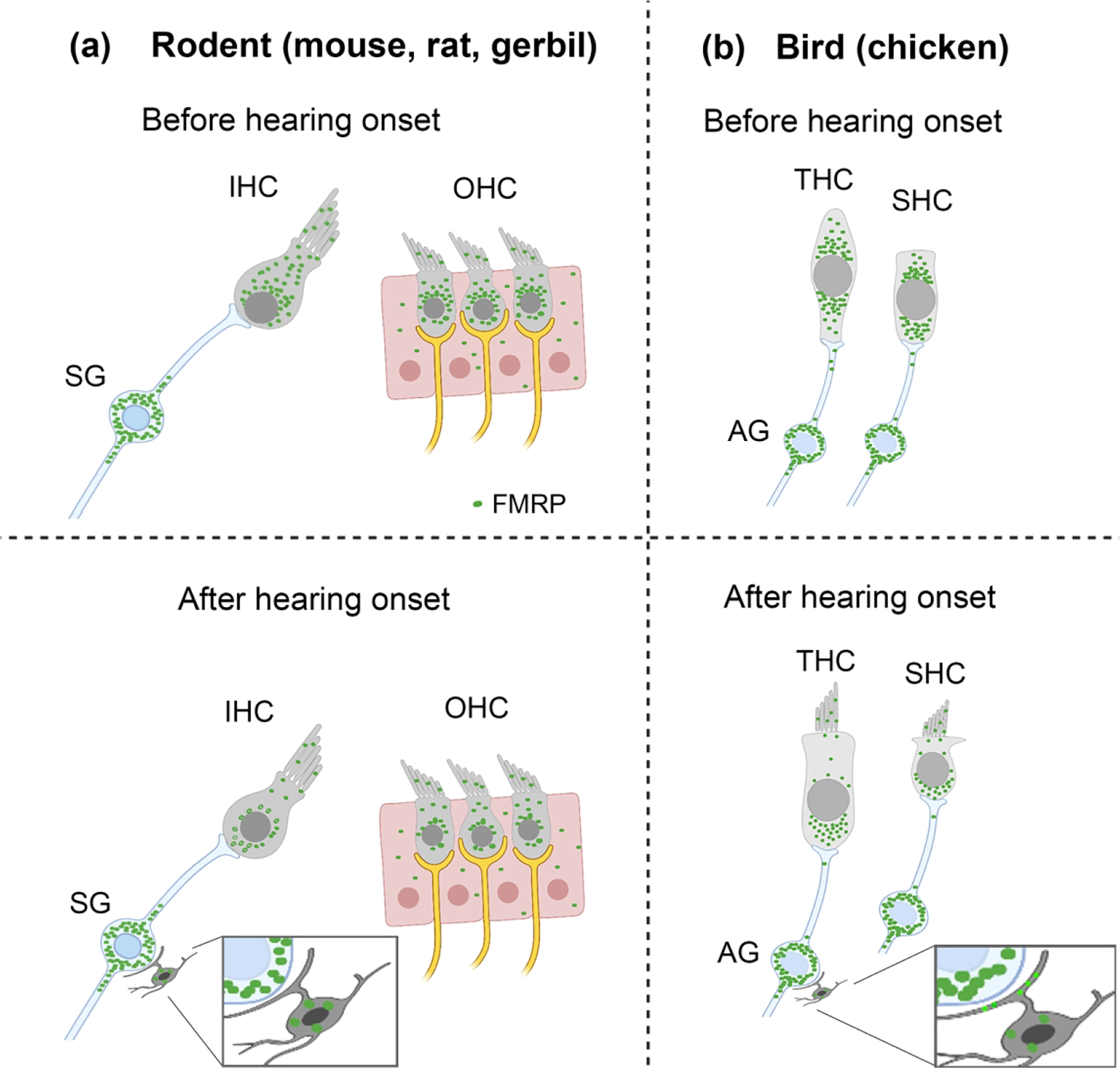
Figure 16.

Schematic drawings of FMRP profile in the cochlea. (a) FMRP expression pattern in the organ of Corti and spiral ganglion (SG) in rodents before and after hearing onset. (b) FMRP expression pattern in the chicken hair cells and auditory ganglion (AG). Green particles indicate the presence of FMRP immunoreactivity.
