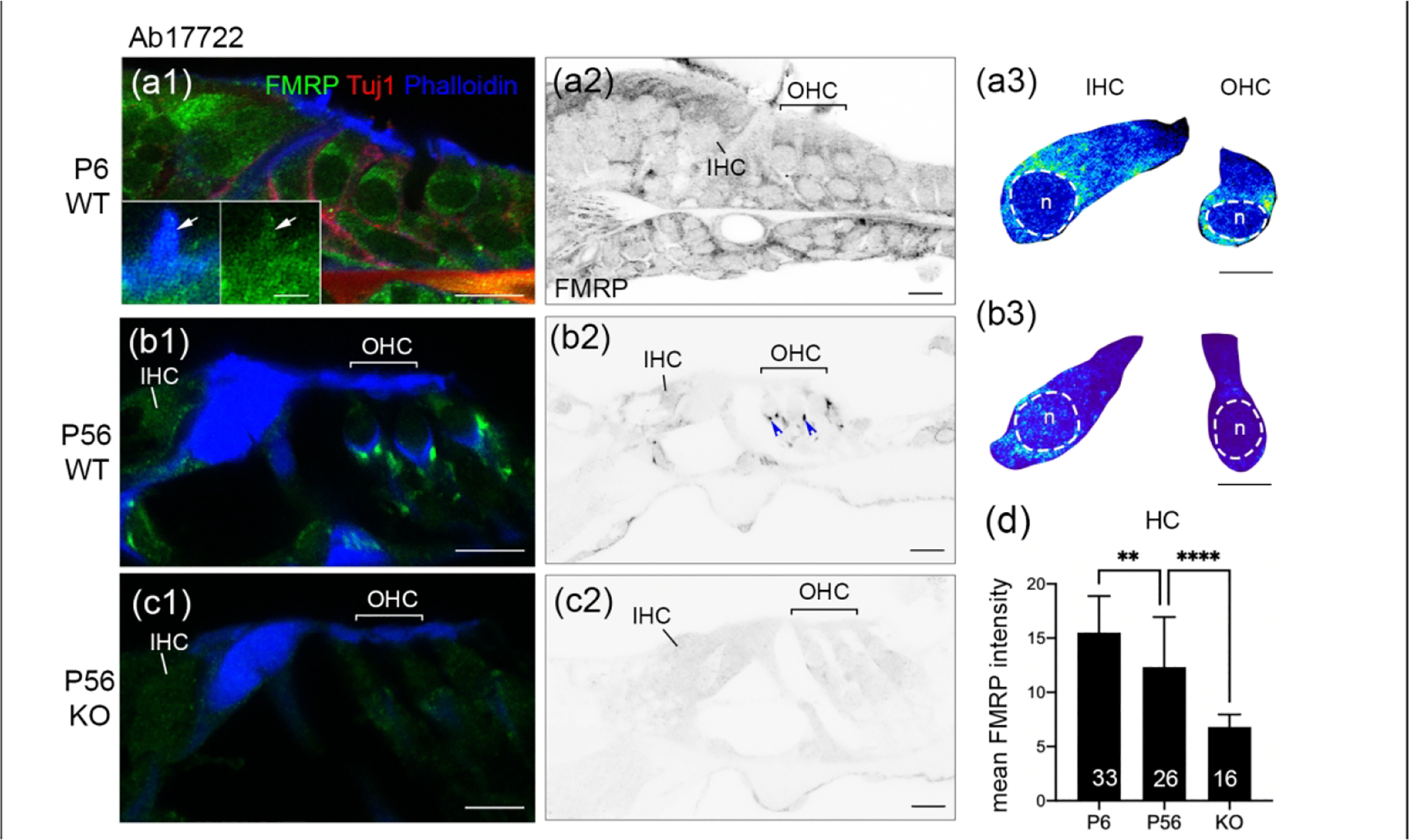
Figure 3.

FMRP_Ab17722 immunostaining in the mouse organ of Corti during development. (a–c) Ab17722 immunoreactivity on midmodiolar sections from WT P6 (a1–a2), WT P56 (b1–b2), and Fmr1 KO P56 (c1–c2) mice. Sections were double stained for Tuj1 immunoreactivity and Phalloidin counterstain, a marker for hair bundles. Insets in a1 are magnifications of a hair cell bundle (white arrow). a3 and b3 are heat maps of FMRP intensity in inner hair cells (IHCs) and outer hair cells (OHCs) at P6 and P56, respectively. Warmer colors represent higher intensities of FMRP immunoreactivity than colder colors. At each age, the same color scale was applied to IHCs and OHCs. The nuclei of hair cells are outlined with dashed circles and labeled with “n”. (d) Mean intensity of FMRP_Ab17722 in hair cells. The number of hair cells measured is indicated in the corresponding bar. Data were analyzed by one-way ANOVA followed by Tukey’s multiple comparisons: F (2,72) = 32.11; p < 0.0001. **p<0.01; ****p<0.0001. Scale bars = 10 μm in a1, a2, b1, b2, c1, c2; 2 μm in inset of a1; 5 μm in a3, b3.
