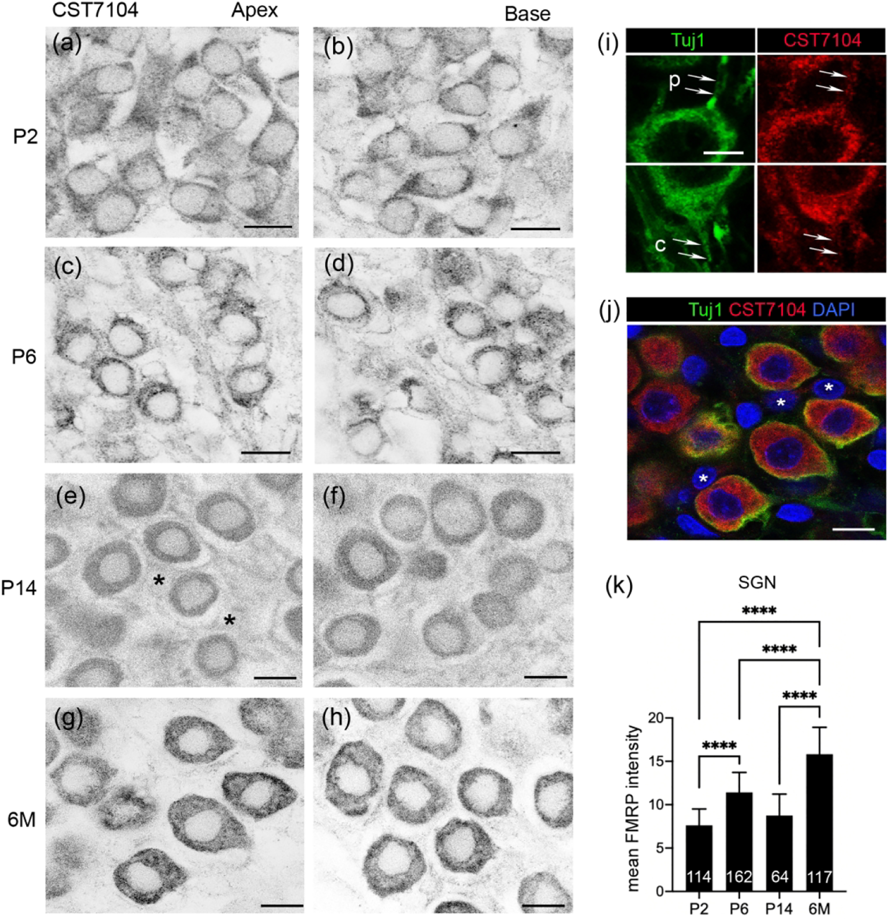
Figure 7.

FMRP_CST7104 immunostaining in the rat spiral ganglion (SG) during development. (a–h) CST7104 immunoreactivity at P2 (a–b), P6 (c–d), P14 (e–f), and 6 months (g–h). The left (a, c, e, g) and right (b, d, f, h) columns were taken from the apex and base of the SG, respectively. Asterisks in (e) indicate the nuclei of satellite glial cells. (i–j) Confocal images of double immunostaining of CST7104 and Tuj1 in the SG of a 6-month-old rat. Arrows point to the peripheral (p) and central (c) processes of a SG neuron. (j) Triple staining for FMRP and Tuj1 immunoreactivities as well as DAPI counterstain. The same region as in (g). White asterisks indicate the nuclei of satellite glial cells. (k) Mean intensity of FMRP_CST7104 in rat SG neurons. The number of neurons measured at each age is indicated in the corresponding bar. Data were analyzed by one-way ANOVA followed by Tukey’s multiple comparisons: F (3, 453) = 251.5; p < 0.0001. ****p<0.0001. Scale bars = 10 μm in a–h, j; 5 μm in i.
